Nanoscale imaging of clinical specimens using pathology-optimized expansion microscopy
Zhao, Y. *, Bucur, O. *, Irshad, H. et al. Nanoscale imaging of clinical specimens using pathology-optimized expansion microscopy. Nat Biotechnol 35, 757–764 (2017). https://doi.org/10.1038/nbt.3892; * equal contribution
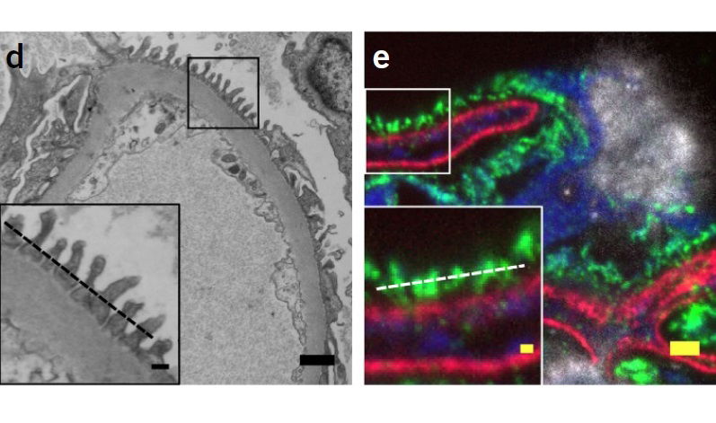
Expansion microscopy (ExM), a method for improving the resolution of light microscopy by physically expanding a specimen, has not been applied to clinical tissue samples. Here we report a clinically optimized form of ExM that supports nanoscale imaging of human tissue specimens that have been fixed with formalin, embedded in paraffin, stained with hematoxylin and eosin, and/or fresh frozen. The method, which we call expansion pathology (ExPath), converts clinical samples into an ExM-compatible state, then applies an ExM protocol with protein anchoring and mechanical homogenization steps optimized for clinical samples. ExPath enables ∼70-nm-resolution imaging of diverse biomolecules in intact tissues using conventional diffraction-limited microscopes and standard antibody and fluorescent DNA in situ hybridization reagents. We use ExPath for optical diagnosis of kidney minimal-change disease, a process that previously required electron microscopy, and we demonstrate high-fidelity computational discrimination between early breast neoplastic lesions for which pathologists often disagree in classification. ExPath may enable the routine use of nanoscale imaging in pathology and clinical research.